Two important internal features in gemstones are inclusions and fissures. Inclusions are an important source of information in the gemological evaluation process. They can often provide indications about geographic origin (see, e.g., S. Saeseaw et al., “Three-phase inclusions in emerald and their impact on origin determination,” Summer 2014 G&G, pp. 114–132); growth conditions (e.g., A. Cheilletz et al., “Time-pressure and temperature constraints on the formation of Colombian emeralds: An 40Ar/39Ar laser microprobe and fluid inclusion study,” Economic Geology, Vol. 89, No. 2, 1994, pp. 361–380; J.G. Toloza et al., “Similarities and differences between fluid inclusions hosted by Colombian emeralds,” Special Issue on the 15th IAGOD Symposium, 2018, pp. 166–167); natural or synthetic origin (e.g., N.D. Renfro et al., “Chart: Inclusions in natural, synthetic, and treated emerald,” Winter 2016 G&G, pp. 402–403); and whether the stone has been treated to improve clarity.
Fissures are openings in the stones, and those “empty” spaces affect clarity in a negative way. Therefore, gemstones are treated to fill those gaps with different substances. Fillers can generate physical phenomena that resemble natural inclusions and may be misleading to the untrained eye.
Emeralds are most commonly enhanced with fissure filling. Fillers in emerald are identified by looking at the flash of color (blue, violet, orange, and yellowish) shown when the transmitted light of the microscope hits the inclusions or with the use of FTIR or Raman spectroscopy.
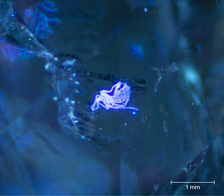
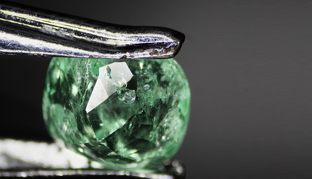
Recently the authors identified a man-made “inclusion” in a 1.15 ct cushion-cut Colombian emerald (figure 1) measuring 8.2 × 7.3 × 3.1 mm. It displayed a typical yellow flash most commonly seen in emeralds filled with liquid resin, which was later corroborated by FTIR spectroscopy. The “inclusion” was barely visible and almost transparent under regular microscope light sources (transmitted and reflected). Therefore, a 365 nm UV light was employed to check whether the foreign object fluoresced, and in fact it did (figure 2).
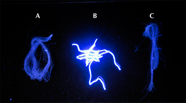
In the authors’ opinion, the fiber could only have come from one of three sources: a cotton handkerchief, a microfiber jewelry cloth, or the net used during the immersion of the stones in the enhancement container of the oiling machine. The first two would imply that a piece of fiber got stuck to the surface of the stone before the enhancement procedure took place, which is highly unlikely. The third source seemed the most plausible. In order to test that hypothesis, three strains of fibers were collected and compared using Raman spectroscopy with a Horiba HR Evolution with the laser operating at 532 nm. Although the spectra showed peaks in the 500 to 1500 cm–1 range, they were not specific for either of the fibers. Using the Olympus BX 41 microscope at 1000×, the diameter of the fiber was measured, and there was consistency with the diameter of the fiber collected from the net, about 50 μm. A 365 nm UV light source was also used to differentiate among them. A clear difference was evident between the nylon fiber from the net and the other two fibers (figure 3). This second test corroborated the authors’ hypothesis.
Article Credit: GIA